Abstract
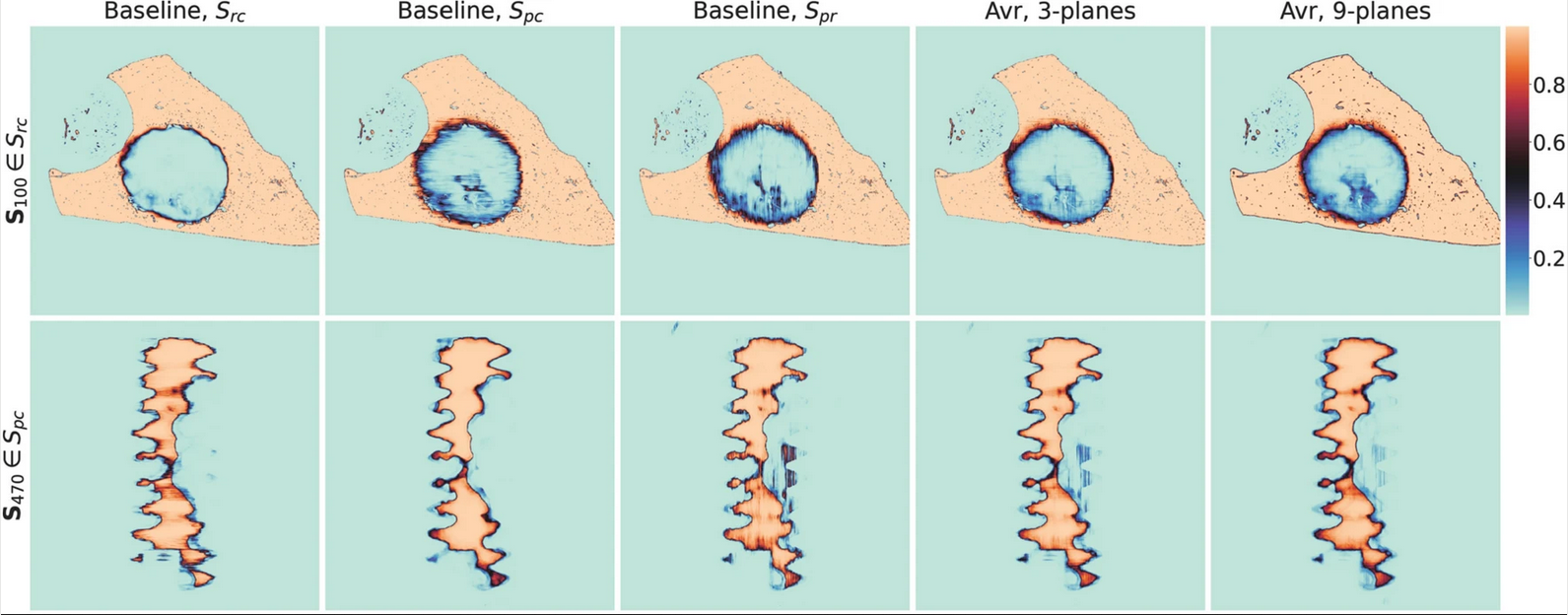 |
Highly accurate segmentation of large 3D volumes is a demanding task. Challenging applications like the segmentation of synchrotron radiation microtomograms (SRμCT) at high-resolution, which suffer from low contrast, high spatial variability and measurement artifacts, readily exceed the capacities of conventional segmentation methods, including the manual segmentation by human experts. The quantitative characterization of the osseointegration and spatio-temporal biodegradation process of bone implants requires reliable, and very precise segmentation. We investigated the scaling of 2D U-net for high resolution grayscale volumes by three crucial model hyper-parameters (i.e., the model width, depth, and input size). To leverage the 3D information of high-resolution SRμCT, common three axes prediction fusing is extended, investigating the effect of adding more than three axes prediction. In a systematic evaluation we compare the performance of scaling the U-net by intersection over union (IoU) and quantitative measurements of osseointegration and degradation parameters. Overall, we observe that a compound scaling of the U-net and multi-axes prediction fusing with soft voting yields the highest IoU for the class “degradation layer”. Finally, the quantitative analysis showed that the parameters calculated with model segmentation deviated less from the high quality results than those obtained by a semi-automatic segmentation method. |
Ivo M. Baltruschat et al., Scaling the U-net: segmentation of biodegradable bone implants in high-resolution synchrotron radiation microtomograms, Sci Rep